

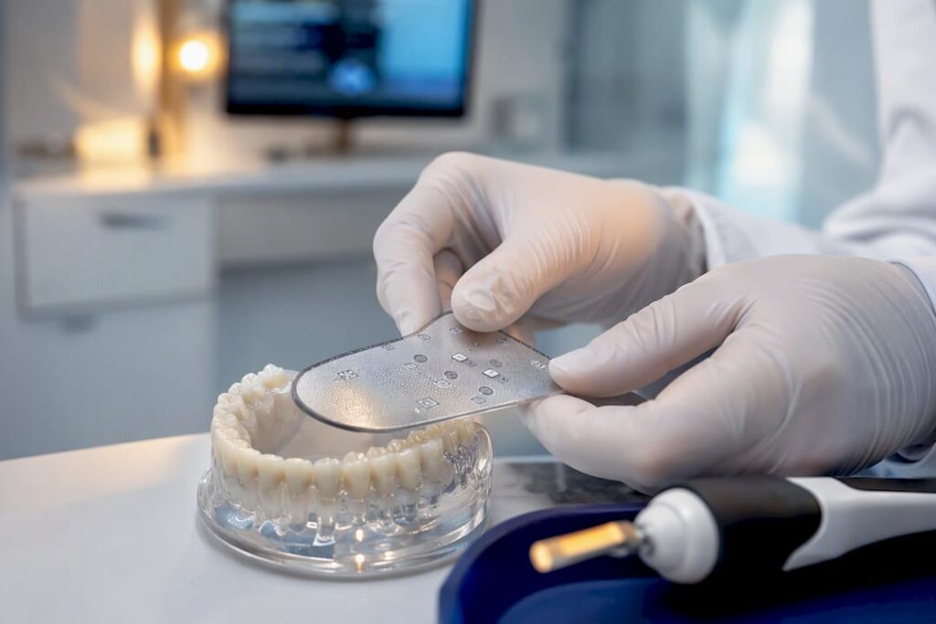
Digital bite analysis uses a thin intraoral sensor and paired software to record the exact timing and relative force of every tooth contact when you bite down, generating a map that shows where your bite lands hardest and fastest. The chief clinical payoff is objective, repeatable data. Instead of guessing whether a filling feels “a little high,” clinicians using systems like T-Scan can pinpoint the exact millisecond and location of an early contact, adjust it, and confirm the fix on the spot.
TL;DR:
- Digital bite analysis provides precise timing and force data, which helps identify problematic contacts that traditional articulating paper cannot reveal.
- Calibration, sensor wear, and proper technique are essential for reliable measurements, and clinicians should perform multiple recordings and regular checks.
- Combining digital analysis with articulating paper enhances diagnostic accuracy by confirming contact location and force distribution.
- Validated systems yield consistent data for treatment planning but should not replace clinical judgment or follow-up verification protocols.
- Objectively measuring bite forces can reduce remakes and adjustments, especially in restorative, TMJ, orthodontic, or bruxism treatments, when used with scrutiny.
Table of Contents
- What Digital Bite Analysis Measures That Articulating Paper Cannot
- How the Technology Works, From Sensor to Screen
- Where Digital Bite Analysis Changes Treatment Decisions
- How Accurate Is Digital Bite Analysis, and Where Does It Fall Short
- What Happens During a Digital Bite Analysis Appointment
- How to Choose a Clinic for Digital Occlusal Analysis
- How Bay Area Dental, Airway & Sleep Uses Occlusal Data
- Digital Bite Analysis Inside a CAD/CAM Workflow
- Reading the Force Map: How Clinicians Turn Data Into Decisions
- What Training Digital Bite Analysis Actually Requires
- Keeping the Equipment Accurate: Maintenance and Calibration
- Why the Real Value Isn’t the Technology, It’s the Discipline Around It
- Ready to Get Your Bite Measured Objectively
- Sources
What Digital Bite Analysis Measures That Articulating Paper Cannot
Articulating paper has done one job for a century: mark where teeth touch. It’s fast, cheap, and tells you almost nothing about sequence or force. Digital bite analysis fills that gap. The sensor records contact across the entire arch in real time, so a clinician sees not just where teeth meet but which tooth hits first, how hard, and how the load shifts as the jaw closes fully.
That distinction matters for a simple reason: two contacts can look identical on paper and behave completely differently under load.
- Paper answers: “Which teeth are touching?”
- Digital sensors answer: “Which tooth hits first, how much force does it carry, and does the bite settle evenly within a fraction of a second?”
Most clinicians who use sensor-based analysis still keep paper in the room. Paper confirms contact location; the sensor confirms timing and force. Used together, they catch problems that neither tool would flag alone, which is why comprehensive reviews of bite force devices describe sensor systems as adjuncts to traditional exam methods, not replacements for them.
How the Technology Works, From Sensor to Screen
The sensor itself is a horseshoe-shaped film, thinner than a sheet of paper, embedded with a grid of pressure-sensitive points. Three sensor technologies dominate the market, and each makes a different trade-off:
- Piezoresistive sensors (used in T-Scan) change electrical resistance under pressure and offer high-resolution timing data across thousands of contact points per second.
- Piezoelectric sensors generate a voltage proportional to force and tend to excel at capturing rapid, dynamic loading events.
- Thin-film resistive sensors, like those in FlexiForce, are lower-cost and often used in research settings or single-point force measurement rather than full-arch mapping.
A typical recording protocol runs through several bites: a relaxed baseline closure, one or two maximum-intensity bites, lateral and protrusive excursions, and then a repeat set after any adjustment. Each recording produces three readable outputs: a force-over-time graph showing how quickly the bite stabilizes, a color-coded pressure map of the arch, and a numeric percentage breakdown of force distribution between left and right sides.
Quick fact: Validation research on modern occlusal-force devices finds these systems can produce reliable data for tracking changes over time, though absolute force readings vary by device and depend heavily on proper calibration.
Where Digital Bite Analysis Changes Treatment Decisions
Objective occlusal data earns its place in several distinct clinical situations, each with its own question to answer.
- Restorative dentistry. Does a new crown or filling carry more load than the surrounding teeth? Digital analysis flags high spots in seconds instead of relying on a patient’s subjective “it feels off.”
- TMJ and TMD evaluation. Is the jaw closing evenly, or is one side absorbing a disproportionate share of force? Tekscan positions T-Scan as an objective measurement and verification tool here, not a standalone diagnostic for joint disorders.
- Orthodontic finishing. Are the back teeth sharing load appropriately once braces or aligners come off?
- Bruxism management. Where is destructive force concentrated during clenching, and has a night guard actually redistributed it?
- Treatment verification. Did the adjustment actually work, or does it just feel better today?
The measurable benefit shows up in fewer remakes and adjustments done by feel. One common scenario: a patient with chronic jaw soreness on one side gets a dynamic recording that shows a single molar taking the first and heaviest contact on closure, a fraction of a second before the rest of the arch settles. A small equilibration on that tooth, verified with a follow-up recording, resolves the soreness within days rather than requiring repeated guesswork visits.
Pro Tip: Ask to see your own force map before and after any adjustment. If a clinic can’t show you a side-by-side comparison, they likely aren’t using the verification loop that makes this technology worth the appointment time.
How Accurate Is Digital Bite Analysis, and Where Does It Fall Short
The evidence supports cautious confidence, not blind trust. Validation studies on modern occlusal-force devices show acceptable reliability for tracking progression and comparing before/after states, which is exactly how most clinicians use the technology day to day. A more recent validation of a piezoresistive gnathodynamometer found excellent reproducibility as a research instrument, though the authors note more work is needed before broad claims can be made across varied patient groups.
Limitations are real and worth naming plainly:
- Sensor films wear with repeated use, and thin-film fatigue can degrade accuracy over time.
- Calibration drift affects absolute force readings more than relative timing data.
- Intraoral alignment matters; a sensor seated slightly off can skew results.
- Absolute force values vary between device types, making cross-brand comparisons unreliable.
Sensor wear and thin-film fatigue reduce repeatability over many uses, which is why clinical labs typically replace sensors on a set schedule and run calibration checks to preserve measurement fidelity.
The mitigation is straightforward: take multiple recordings per visit, calibrate on schedule, and pair sensor data with articulating paper rather than relying on either tool alone.
What Happens During a Digital Bite Analysis Appointment
The procedure itself is short and low-drama. A clinician explains what the sensor will record, seats the horseshoe-shaped sensor between your teeth, and asks you to bite naturally, then firmly, then move through a few side-to-side and forward positions. Recordings take only a few minutes, and the sensor itself is disposable, so nothing carries over from patient to patient.
- Explanation of what’s being measured and why
- Baseline and maximum-force bite recordings
- Adjustments, if the data shows a problem worth correcting
- A verification recording to confirm the fix worked
Cost for a standalone bite analysis session typically varies and may be bundled into other dental appointments rather than billed separately. Insurance coverage varies widely by plan and diagnosis code, so it’s worth confirming coverage with your insurer before the visit rather than assuming it’s included.
How to Choose a Clinic for Digital Occlusal Analysis
Not every practice that owns a sensor system uses it well. The equipment is only as good as the clinician reading the output.
- Ask how often they use it. A device that gathers dust in a drawer produces worse data than one used weekly.
- Request to see anonymized before/after records. A clinician confident in their workflow will have examples ready.
- Ask about their verification protocol. Do they re-record after every adjustment, or trust the first pass?
- Confirm whether they combine digital analysis with articulating paper. The strongest practices use both.
- Ask about follow-up. Occlusion can shift over weeks; a single recording without a follow-up plan tells an incomplete story.
Pro Tip: A red flag worth walking away from: any clinician who promises a permanent, one-visit fix for TMJ pain based on a single bite recording. Objective data is powerful, but joint disorders are rarely solved by one adjustment.
How Bay Area Dental, Airway & Sleep Uses Occlusal Data
Bay Area Dental, Airway & Sleep integrates digital bite analysis into multidisciplinary care rather than treating it as an isolated test. For patients also working through airway therapy or myofunctional therapy, an uneven bite can complicate breathing patterns and tongue posture during sleep, so occlusal data gets read alongside those findings rather than in isolation. [Author credentials, internal case outcomes, and patient testimonials to be added.]
Digital Bite Analysis Inside a CAD/CAM Workflow
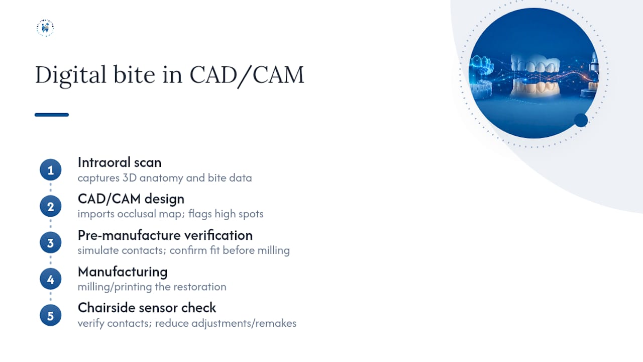
Digital occlusal data doesn’t live in isolation anymore. Most modern restorative workflows already run through an intraoral scanner and CAD/CAM design software, and bite force data slots directly into that pipeline. When a lab designs a crown, bridge, or aligner sequence digitally, the occlusal contact data from a sensor recording can confirm that the digital design will actually perform correctly once seated, before a single unit of material gets milled.
A 2024 evaluation of digital and virtual bite registration methods found that digital approaches generally beat older analogue registration techniques on accuracy, speed, and integration into clinical workflows. That matters practically: a patient getting a same-day crown no longer needs a separate physical bite registration step that risks distortion in transit or during pouring. The scan, the design, and the occlusal verification happen in one connected digital chain.
This integration also changes how adjustments get made. Instead of a lab technician guessing at contact points from a plaster model, the design software can flag potential high spots before manufacturing, and the finished restoration can be checked with a sensor recording chairside. For orthodontic aligner sequences, some workflows now use baseline occlusal maps to inform how tooth movement plans account for existing force imbalances, rather than treating the bite as a static target to hit at the end of treatment.
The practical upshot for patients: fewer adjustment visits, fewer remakes, and a restoration more likely to feel correct on the first fitting.
Reading the Force Map: How Clinicians Turn Data Into Decisions
A force map without interpretation is just a colorful picture. The skill lies in reading what the timing and distribution actually mean for treatment.
The first thing a clinician looks at isn’t where the heaviest contact sits. It’s the sequence. A healthy closure typically shows contacts landing close together in time, with force distributing relatively evenly across both sides within a fraction of a second. A single tooth lighting up red well before the rest of the arch, even if it’s not the highest-force point, often points to the actual problem tooth. That’s counterintuitive to patients who assume the darkest red spot on the map is automatically the culprit.
Left/right force percentage matters almost as much as timing. A significant and consistent imbalance, especially one that shows up across multiple recordings rather than a single bite, suggests a structural issue worth investigating rather than a fluke of that particular closure. Clinicians generally treat a single recording with skepticism and want to see the pattern repeat across two or three bites before acting on it.
The decision to adjust versus monitor comes down to correlating the data with the patient’s actual symptoms. A minor imbalance on someone with no complaints usually gets logged and watched, not touched. The same imbalance in a patient reporting jaw fatigue or morning headaches becomes a target for equilibration, verified immediately with a follow-up recording. The data guides the decision, but it never replaces the clinical exam and the patient’s own reported symptoms.
What Training Digital Bite Analysis Actually Requires
Owning the equipment and knowing how to read it are two different skill sets, and the gap between them explains why results vary so much between practices. The hardware itself isn’t hard to operate. Seating a sensor and running a recording takes a fraction of the training that interpreting the output does.
Real competency comes from repetition. Clinicians who use sensor-based analysis weekly develop an intuitive sense for what a normal timing sequence looks like versus a problematic one, the same way an experienced radiologist reads a scan faster and more accurately than someone who sees one a month. Vendor training programs for systems like T-Scan typically cover software navigation and basic interpretation, but building real diagnostic judgment takes ongoing case volume.
There’s also a softer skill involved: knowing when the data doesn’t tell the full story. A clinician trained only on the software risks over-treating a bite that looks imbalanced on screen but causes the patient no functional problem. The better-trained clinician cross-references the map against the physical exam, the patient’s symptom history, and, increasingly, findings from articulating paper, before deciding whether to touch anything at all.
Continuing education courses through dental schools and manufacturer-run programs exist specifically to close this gap, and practices that invest in ongoing training tend to produce more consistent, defensible treatment decisions.
Keeping the Equipment Accurate: Maintenance and Calibration
A sensor system is only trustworthy if it’s maintained on a schedule, and this is where a surprising number of practices cut corners. The sensors themselves are consumable. Each one is designed for single use or a limited number of recordings before the thin-film material fatigues and starts producing less reliable readings, which is a documented factor in why measurement fidelity degrades over repeated use.
Calibration is a separate issue from sensor wear. The handpiece and software need periodic calibration checks against known reference forces to keep absolute readings accurate, even though relative timing data tends to hold up better between calibrations than absolute force numbers do. Practices that skip this step can still get useful comparative data (this bite versus that bite) while losing confidence in the precise force numbers displayed.
Storage and handling matter more than most patients would guess. Sensors are sensitive to bending and moisture, and mishandling between patients can introduce inconsistency that has nothing to do with the patient’s actual bite. Well-run practices log sensor replacement dates and calibration checks the same way they’d log sterilization cycles, treating it as a routine maintenance task rather than an occasional afterthought.
The takeaway for patients: a practice that treats calibration as routine, not an afterthought, is one whose numbers you can trust further.
Why the Real Value Isn’t the Technology, It’s the Discipline Around It
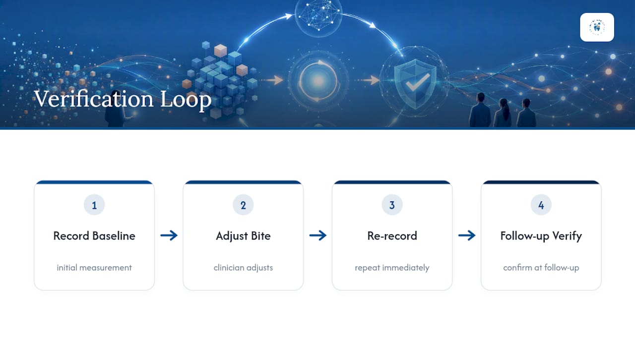
The conventional pitch for digital bite analysis leans hard on the gadget: sensors, software, colorful maps. That framing undersells what actually makes the data useful. The evidence consistently points to something less flashy: the verification loop. Record a baseline, make an adjustment, re-record immediately, and check again at a follow-up visit. Skip that loop and even the most advanced piezoresistive sensor produces a single snapshot with no way to confirm it worked.
Where conventional marketing overpromises is treating a bite map as a diagnosis unto itself. It’s not. Tekscan itself frames T-Scan as a measurement and verification tool, not a standalone answer for TMJ disorders, and the peer-reviewed literature backs that caution. A red spot on a screen still needs a clinician who correlates it with symptoms, a physical exam, and a bite paper mark.
If you take one thing from this: prioritize a clinic’s follow-up habits over its equipment brand. A practice with an older system that re-records after every adjustment will likely serve you better than one with the newest sensor and no verification protocol at all.
— Admin
Ready to Get Your Bite Measured Objectively
If chronic jaw soreness, a restoration that never quite feels right, or unexplained headaches have you wondering whether your bite is the culprit, a data-backed answer beats another round of guesswork. Bay Area Dental, Airway & Sleep in Fremont pairs digital bite analysis with occlusal adjustment and airway assessment, so a force imbalance doesn’t get treated as an isolated dental issue when it might connect to breathing or sleep patterns too.

A new patient evaluation typically starts with a conversation about your symptoms, moves into a bite recording session, and ends with a clear plan, whether that means a targeted adjustment, a referral into airway or myofunctional therapy, or simply confirmation that nothing needs to change. You can review services and book an evaluation directly through the practice’s site to get a bite analysis scheduled and find out what your own force map actually shows.
This article is general information, not a substitute for advice from a qualified doctor. Consult a qualified healthcare professional about your own circumstances before acting on anything here.