

Minimally invasive dentistry treats decay as a disease to manage, not just a hole to fill, preserving as much healthy tooth structure as possible. It relies on early detection, risk-based monitoring, and techniques like selective caries removal, resin infiltration, and silver diamine fluoride instead of automatic drilling. Dentists use it for early lesions, pediatric patients, and high-risk mouths where watching and arresting decay beats cutting it out. Groups like the AAPD back this approach, but only when paired with real follow-up care.
TL;DR:
- Minimal intervention prioritizes monitoring decay progression with risk assessments over immediate drilling, especially in early or non-cavitated lesions.
- Techniques like selective caries removal, resin infiltration, and silver diamine fluoride enable preservation of healthy tooth structure and reduce patient discomfort.
- Modern materials such as glass ionomers and bioactive cements support conservative treatments by promoting remineralization and bonding without extensive removal.
- Follow-up and risk-based scheduling are essential, with intervals tailored to individual hygiene, lesion activity, and patient age, not a standard six-month cycle.
- Professional guidelines endorse minimally invasive dentistry as part of disease management, emphasizing active monitoring and appropriate treatment rather than default restoration.
Table of Contents
- The Philosophy Behind Minimally Invasive Dentistry
- Gentle Dental Techniques That Preserve Tooth Structure
- Materials That Make Conservative Dentistry Possible
- How Dentists Plan and Monitor Minimally Invasive Care
- Weighing the Benefits Against the Real Limitations
- What the AAPD and ADA Actually Recommend
- How This Looks in Practice at a Modern Dental Office
- Why the “Just Fill It” Mindset Is Losing Ground
- Finding a Dentist Who Practices This Approach
- Sources
The Philosophy Behind Minimally Invasive Dentistry
Every drill cut into a tooth is permanent. That single fact drives the entire logic of minimally invasive dentistry: once healthy enamel and dentin are gone, they don’t come back, so the goal shifts from “fix the cavity” to “keep the tooth alive with as much of its original structure intact as possible.” This isn’t a trend or a marketing term. It reflects a documented shift away from routine removal and replacement of tooth structure toward preventing disease from progressing in the first place.
Dentistry researchers sometimes describe the clinical foundation of this approach as a “golden triangle” connecting three disciplines: understanding how decay actually spreads through dentin (histopathology), choosing materials that bond and release protective ions (biomaterials), and applying gentle, precise clinical handling. Miss any one leg of that triangle and the whole approach falls apart. A dentist can have the best bioactive filling material available, but it means nothing without the tactile skill to know when to stop removing tooth structure.
This translates into a few practical shifts in how care gets planned:
- Decay gets tracked and risk-scored, not just discovered and drilled.
- Prevention and interception (catching a lesion before it cavitates) take priority over restoration.
- Recall intervals get tied to individual risk, not a blanket six-month rule for everyone.
- Treatment decisions weigh long-term tooth survival, not just closing the immediate hole.
A low-risk adult with excellent hygiene and no new cavities in a decade might go a full year between checkups. A child with multiple active white-spot lesions might need monitoring every three months with fluoride varnish and sealants layered in between. Same philosophy, very different schedules, because ongoing surveillance is what makes conservative treatment safe.
Gentle Dental Techniques That Preserve Tooth Structure
Several procedures now let dentists treat decay without the traditional drill-and-fill sequence. Each targets a different stage of disease, and understanding which technique fits which situation helps you ask better questions at your next visit.
- Selective (partial) caries removal. Instead of excavating until dentin feels rock hard everywhere, the dentist removes only the soft, infected outer layer and leaves firmer, remineralizable dentin near the pulp. Practitioners judge this largely by feel. Clinical research on tactile assessment describes dentin as “leathery” versus “hard,” and stopping at leathery dentin near the pulp chamber avoids exposing the nerve unnecessarily. This single technique is part of why contemporary caries management increasingly favors partial removal over complete excavation.
- Resin infiltration. For a lesion that hasn’t cavitated yet, often on a tooth’s proximal (side) surface, the dentist etches the enamel, applies a low-viscosity resin that seeps into the porous decayed area, then cures it with light. No drilling at all. It’s especially effective for orthodontic patients who develop white-spot lesions under brackets.
- Silver diamine fluoride (SDF). A liquid brushed directly onto active decay that arrests the lesion chemically rather than removing it. It’s fast, painless, and needs no anesthesia or drilling, which makes it valuable for toddlers, patients with dental anxiety, or anyone who can’t tolerate a longer procedure. The trade-off: it permanently stains treated decay a dark brown to black, so it’s best used on back teeth or baby teeth rather than a front incisor.
- The Hall technique. Used mainly on primary molars, this seals a decayed baby tooth under a preformed metal crown without any local anesthesia, numbing, or removal of decay. The seal itself starves the bacteria of the nutrients they need to keep progressing.
- Interim therapeutic restorations (ITR). A temporary but often long-lasting glass ionomer filling placed with minimal or no drilling, frequently used for young children or patients who need decay controlled before more definitive work.
- Minimally invasive removal tools. Air abrasion (a fine particle spray), smart burs that change resistance based on tissue hardness, and dental lasers all let the dentist remove decay with more precision and less vibration than a traditional handpiece.
Pro Tip: If your dentist mentions SDF for a child’s molar, ask specifically whether the treated tooth will show visible staining and where. Staining on a back molar is rarely noticeable when your child smiles or talks.
Materials That Make Conservative Dentistry Possible
None of these techniques would work without a parallel revolution in dental materials. Older amalgam fillings needed mechanical undercuts carved into the tooth to stay in place, which meant removing healthy structure just to hold the filling. Modern adhesive materials bond directly to tooth surfaces, which is the material science that quietly makes minimally invasive dental treatments possible at all.
- Glass ionomer cement (GIC) and resin-modified glass ionomer (RMGIC): These release fluoride continuously, actively supporting remineralization in the surrounding tooth. That dual role, restoring form while fighting future decay, makes them a natural fit for high-caries-risk patients and pediatric cases, as reviews of biomaterial strategies point out.
- Resin composites and adhesive bonding systems: Because these materials chemically bond to enamel and dentin, the cavity prep can be conservative rather than shaped for mechanical retention.
- Biodentine, MTA, and other bioactive substitutes: These calcium-silicate materials sit close to an exposed or near-exposed pulp and actively encourage the tooth to lay down new dentin, buying time and sometimes avoiding root canal treatment entirely.
None of this is a free lunch. Glass ionomers wear faster than composite and aren’t the first choice for a visible front tooth that needs to look flawless. Composite requires a dry field to bond properly, which is harder to achieve in a squirming toddler. There are still situations, large lesions, structurally compromised teeth, cases with poor moisture control, where a conventional restoration remains the more reliable option.
How Dentists Plan and Monitor Minimally Invasive Care
Good minimally invasive dental treatment starts with an honest risk assessment, not a treatment plan. Dentists typically classify patients as low, moderate, or high caries risk using diet, fluoride exposure, saliva flow, and past cavity history, then layer diagnostic tools on top: visual and tactile exam, radiographs, transillumination (shining light through a tooth to spot hidden decay), and increasingly 3D imaging for a clearer picture of lesion depth before committing to any procedure.
Care then gets sequenced rather than delivered all at once:
- Urgent phase: Address pain or infection first, always.
- Control phase: Arrest active decay with SDF, fluoride varnish, or diet and hygiene changes.
- Definitive phase: Restore what genuinely needs restoring, once the mouth is stable.
- Maintenance phase: Monitor arrested or borderline lesions at intervals matched to risk level.
That staging matters because it stops a dentist from restoring something today that might have resolved on its own with fluoride and time. A shallow, non-cavitated lesion caught on a radiograph doesn’t need a filling next week. It needs a recheck in three to six months to see whether it’s progressing or arresting. Red flags that override the wait-and-watch approach include visible cavitation, pain, radiographic evidence the lesion has reached the pulp, or a patient who simply can’t maintain the hygiene the conservative plan depends on. Shared decision-making runs through all of it: a dentist explaining that a watched lesion carries some risk of progressing, weighed against the certainty of removing healthy tooth structure today, is the conversation that makes minimally invasive dentistry work in practice rather than just on paper.
Weighing the Benefits Against the Real Limitations
The upside is straightforward: less tooth structure removed, lower risk of exposing the nerve, less discomfort during treatment, and procedures that work well even for toddlers and anxious patients who couldn’t tolerate a traditional drill session.
The limitations deserve equal airtime, because glossing over them undercuts trust:
- SDF’s dark staining is permanent on the treated area and not something that fades.
- Larger or structurally compromised lesions still need conventional restoration or crowns.
- Evidence quality varies by technique. Some, like resin infiltration and SDF, have solid backing; others are newer and still building a track record.
- Billing structures in general still reward procedure-based work over prevention or monitoring, which can create friction when a conservative, watch-and-treat plan takes multiple short visits instead of one filling.
Pro Tip: Ask your dentist three questions before agreeing to any plan: What happens if we watch this lesion instead of treating it now? What does success look like at the next visit? And how often will you need to see me to monitor it? Those three answers tell you more about the plan’s soundness than any brand name on the material used.
What the AAPD and ADA Actually Recommend
Professional guidance on this topic is more specific, and more cautious, than most patient-facing marketing suggests. The AAPD’s policy on minimally invasive dentistry frames it explicitly as risk-based and family-centered, endorsing fluoride varnish, SDF, sealants, ITR, and the Hall technique as legitimate tools, provided they come with active monitoring rather than a one-and-done mindset.
The ADA’s evidence-based guideline on restorative treatments takes a similar stance, recommending more conservative carious tissue removal over complete excavation and offering conditional (not absolute) recommendations on material selection, meaning the right material still depends on the specific tooth and patient.
A few points worth holding onto:
- Both organizations treat MID as disease management, not a downgrade from “real” dentistry.
- Evidence strength varies: sealants and SDF have strong support; some newer bioactive materials have thinner long-term data.
- Neither body positions MID as a substitute for restorative care when a tooth genuinely needs it. Watching a lesion that should be treated isn’t conservative, it’s negligent.
How This Looks in Practice at a Modern Dental Office
At Bay Area Dental, Airway & Sleep, this approach shows up in the tools used before a single instrument touches a tooth. 3D imaging gives a clearer look at lesion depth, which supports the call to watch a lesion versus treat it now. Laser treatments allow more precise, gentler removal of decay or soft tissue when treatment is needed, and the practice’s airway and myofunctional therapy work reflects the same root-cause philosophy applied beyond just cavities. Communication gets adapted for each patient, including tailored approaches for Deaf patients, because a monitoring-based plan only works if the patient actually understands and returns for it. If you’re weighing a conservative option, a consultation is the place to confirm risk level, review imaging, and agree on a monitoring schedule together, the same process a risk-based scheduling approach depends on in any well-run practice.
Why the “Just Fill It” Mindset Is Losing Ground
The evidence here doesn’t support a blanket rule for every mouth, and it doesn’t support treating minimally invasive dentistry as automatically superior either. What it supports is something less catchy but more honest: the right approach depends entirely on risk level and lesion behavior over time, and that requires a dentist willing to watch and wait when watching and waiting is the smarter bet.
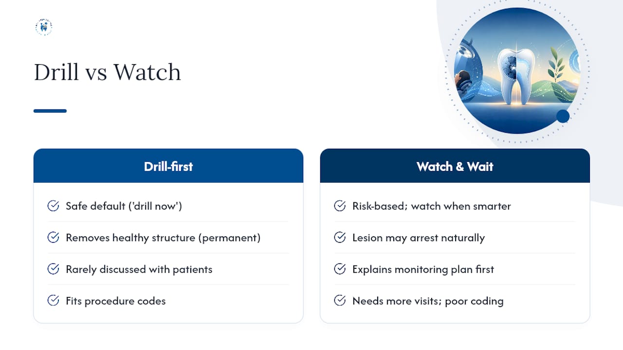
Where conventional advice falls short is in treating “drill it now” as the safe default. It isn’t. Removing healthy tooth structure to treat a lesion that might have arrested on its own carries its own cost, one that’s permanent and rarely discussed with patients in plain terms. At the same time, the billing reality is real: monitoring-based care takes more visits and doesn’t map cleanly onto procedure codes, which quietly pressures some practices toward restoration-first habits regardless of what the evidence says.
If you take one thing from this, prioritize finding a dentist who explains the monitoring plan before proposing the drill. That single habit separates practices actually applying minimally invasive dentistry from ones using the term loosely.
— Admin
Finding a Dentist Who Practices This Approach
A modern dental practice that applies the evidence-based approach treats decay by assessing risk first, treating only what genuinely needs treating, and monitoring the rest with a real follow-up plan instead of a rushed default to the drill. That’s the practical difference for anyone tired of leaving an appointment with more tooth removed than the problem required.

Advanced 3D imaging and laser treatments support the kind of selective, tissue-preserving care this article describes, extending into airway and myofunctional therapy for patients whose dental issues connect to breathing and sleep. Families with young children, anxious patients, and anyone managing early-stage decay may be well suited to this approach, including Deaf patients who can benefit from tailored communication methods. If you want to know whether a conservative, monitoring-based plan fits your situation, schedule a consultation and bring your questions about staging, materials, and recall intervals. That conversation is the actual next step, not another round of research.
This article is general information, not a substitute for advice from a qualified doctor. Consult a qualified healthcare professional about your own circumstances before acting on anything here.