

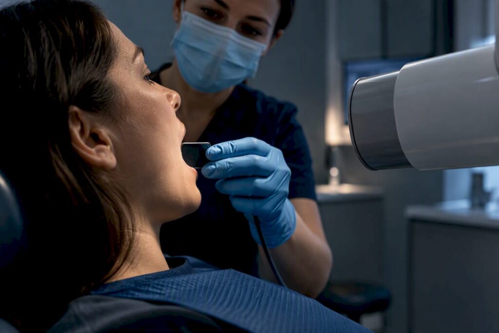
Dental X-rays are low-dose and, when clinically justified and optimized, safe for the overwhelming majority of patients. That verdict rests on three things: the doses involved are tiny compared to everyday radiation, professional bodies like the ADA and FDA require justification before any exposure, and modern digital equipment has slashed doses that were already small to begin with. The exceptions worth knowing about are CBCT scans, which carry a higher dose, and children, who get dedicated protocols.
TL;DR:
- Dental X-ray doses are extremely low, with digital technology reducing radiation by up to 90 percent compared to older film-based systems.
- CBCT scans produce higher doses and should only be used when specific 3D imaging is necessary for complex cases like implant planning or impacted tooth assessment.
- Children and pregnant women require tailored protocols to minimize exposure, with children benefiting from smaller, adjusted doses and pregnancy exams justified only if vital.
- Modern guidelines recommend avoiding routine lead aprons and thyroid collars because precise beam control and digital techniques already limit scatter radiation effectively.
- Patients should actively ask about the necessity, type, and dose-reduction methods of their imaging to ensure exposure is minimized and tailored to their individual needs.
Table of Contents
- Dental X-Ray Safety: The Different Exam Types and Why They Matter
- How Much Radiation Do Dental X-Rays Actually Deliver?
- Who Needs Extra Caution: Children, Pregnancy, and Repeat Exposure
- Modern Dose-Reduction Tools: ALARA, ALADA, and What Changed With Lead Aprons
- CBCT Scans: When a 3D Image Is Actually Worth the Higher Dose
- What You Can Do to Keep Your Own Exposure Low
- How Bay Area Dental, Airway & Sleep Applies Imaging Safety in Practice
- A Note on Trust: Why the Conversation Matters More Than the Machine
- Want an Imaging Approach Built Around ALARA? Here’s What to Expect
- Sources
- FAQ
Dental X-Ray Safety: The Different Exam Types and Why They Matter
Not every dental X-ray carries the same dose or purpose, and knowing the difference helps you understand why your dentist orders one over another.
- Bitewing and periapical X-rays (both intraoral) capture a few teeth at a time to catch cavities between teeth or check root and bone health around a single tooth.
- Panoramic X-rays sweep the entire jaw in one image, useful for spotting impacted wisdom teeth, jaw abnormalities, or planning orthodontic treatment.
- Cephalometric X-rays capture the side profile of the skull and jaw, mostly used in orthodontic planning to measure bite and facial growth.
- CBCT (cone-beam computed tomography) builds a 3D image of teeth, bone, and soft tissue, reserved for complex cases like implant planning or surgical assessment.
Each of these carries a different dose footprint, which is where the numbers actually get reassuring.
How Much Radiation Do Dental X-Rays Actually Deliver?
The doses involved are small enough that most people underestimate just how conservative dental imaging really is. According to dose data from the IAEA, a typical intraoral X-ray delivers between 1 and 8 microsieverts (μSv), a panoramic X-ray runs 4 to 30 μSv, and a cephalometric image sits at just 2 to 3 μSv.
By the numbers: A single panoramic dental X-ray tops out around 30 μSv. A cross-country round-trip flight exposes you to roughly 40 μSv from cosmic radiation at altitude, and average background exposure from soil, air, and cosmic rays runs close to 10 μSv per day in many parts of the country. A chest X-ray delivers around 100 μSv, and a CT scan of the abdomen can reach 10,000 μSv or more.
Digital sensors deserve a lot of the credit here. Cleveland Clinic notes that digital dental X-rays use about 80% to 90% less radiation than the older film-based systems many patients remember from childhood visits. “Effective dose” is the metric that lets researchers compare radiation risk across different body parts and exposure types, and by that measure, dental imaging sits near the bottom of the medical radiation scale, not the top.
Who Needs Extra Caution: Children, Pregnancy, and Repeat Exposure
Radiation risk isn’t uniform across every patient, and dentists adjust their approach accordingly.
- Children’s tissue is more radiosensitive and they have more remaining years of life for any theoretical risk to accumulate, which is why the FDA emphasizes pediatric-specific imaging protocols with reduced exposure settings sized to smaller anatomy.
- Pregnancy doesn’t rule out dental X-rays. When an exam is genuinely necessary, dentists weigh the (very low) risk against the benefit of catching an infection or abscess that could otherwise threaten the pregnancy, and they’ll often delay non-urgent imaging to a more comfortable time.
- There’s no universal schedule for how often X-rays should happen. A patient with a history of cavities and active gum disease needs more frequent monitoring than someone with a clean, stable dental history, so frequency gets individualized rather than applied on autopilot.
Modern Dose-Reduction Tools: ALARA, ALADA, and What Changed With Lead Aprons
Two acronyms drive nearly every imaging decision in a modern dental office: ALARA (As Low As Reasonably Achievable) and its dental-specific cousin ALADA (As Low As Diagnostically Acceptable). Both principles say the same thing in different words: never take and X-ray you don’t need, and when you do need one, use the lowest dose that still produces a diagnostic image.
Here’s what that looks like in the operating room:
- Digital receptors replace film, cutting dose by up to 90% while producing an image that shows up instantly on screen.
- Rectangular collimation narrows the X-ray beam to match the size of the digital sensor, avoiding unnecessary radiation to surrounding tissue.
- Exposure settings are tuned per patient, with pediatric-specific and size-based protocols reducing dose further for smaller patients.
One change surprises a lot of patients: the ADA’s updated guidance says thyroid collars and lead aprons are no longer routinely recommended. Modern beam control is precise enough that scatter radiation to the rest of the body is minimal, and in some cases a poorly placed apron can actually cause image errors that require a retake, which means a second dose instead of avoiding one.
Pro Tip: If your dentist skips the lead apron, that’s not a shortcut. It usually means their equipment and technique already meet the newer, tighter guidelines, and adding a shield wouldn’t meaningfully lower your exposure.
CBCT Scans: When a 3D Image Is Actually Worth the Higher Dose
CBCT is the one dental imaging technology where the dose conversation gets more serious. The FDA is direct about this: CBCT typically delivers more radiation than a standard 2D dental X-ray, and it should only be used when a 2D image genuinely won’t answer the clinical question.
Legitimate reasons for CBCT include:
- Planning dental implant placement, where 3D bone measurements matter
- Assessing complex oral surgery cases, like impacted teeth near nerves
- Select orthodontic cases involving jaw or airway structure
Peer-reviewed dose-optimization research shows that adjusting filtration and kilovoltage settings can cut CBCT dose by 40% to 57% in some units, which is exactly why field-of-view size matters so much. Before a CBCT scan, it’s fair to ask your dentist: is this necessary, would a 2D image suffice, and what field of view will be used? A smaller field of view, targeting only the area in question, keeps dose down.
What You Can Do to Keep Your Own Exposure Low
You have more say in this process than most patients realize.
- Ask directly whether the X-ray is necessary right now, and what specific type is being ordered.
- Request pediatric exposure settings for children if you’re not certain the office defaults to them.
- Offer to bring prior X-rays from a previous dentist. The ADA supports using existing images when they’re diagnostically current, which avoids a repeat exposure entirely.
- Ask what dose-reduction techniques the office uses, such as digital sensors or rectangular collimation, and expect a straight answer.
How Bay Area Dental, Airway & Sleep Applies Imaging Safety in Practice
Some dental practices treat imaging as a diagnostic tool tied to root causes, not a routine box to check. A whole-body approach to airway and sleep dentistry means 3D imaging may be used when it genuinely changes a treatment plan, not by default. The use of laser treatments and minimally invasive technology reflects the philosophy of doing less when less accomplishes the same goal.
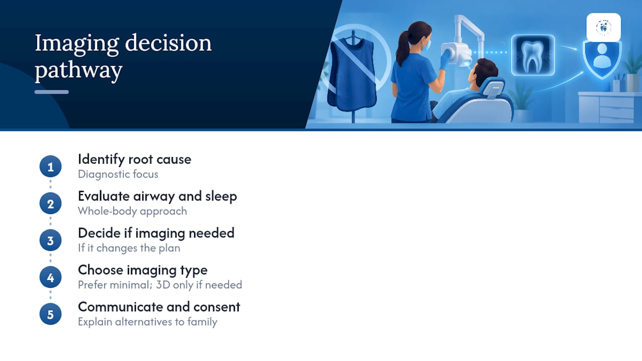
For pediatric patients and families evaluating tongue and lip ties, dental teams often discuss why an image is needed and what alternatives exist before proceeding. A communication-first approach often extends to work with Deaf patients, where clear, accessible explanations of any procedure, imaging included, may be part of standard care.
A Note on Trust: Why the Conversation Matters More Than the Machine
The safety verdict on dental X-rays holds up. Low doses, clear guidelines, and better equipment have made this one of the more conservative corners of medical imaging. What actually protects you isn’t the machine, though. It’s whether your dentist explains why an image is needed and tailors the plan to you rather than defaulting to habit. Ask questions. Expect answers.
— Admin
Want an Imaging Approach Built Around ALARA? Here’s What to Expect
Some dental practices follow ALARA and ALADA principles as a baseline, using optimized digital equipment and size-based protocols rather than one-size-fits-all imaging.

If you’re weighing a first visit or a second opinion on whether an X-ray, or a CBCT scan, is genuinely necessary for your case, bring any prior images from a previous dentist so the team can review them before ordering anything new. Families evaluating pediatric care can look at the practice’s pediatric dental services to see how imaging fits into a broader, child-specific care plan, while adults exploring routine or restorative needs can start with general dentistry services. To schedule a consultation and ask these questions directly, visit Bay Area Dental, Airway & Sleep to book an appointment.
This article is general information, not a substitute for advice from a qualified doctor. Consult a qualified healthcare professional about your own circumstances before acting on anything here.
Sources
- Dental X-rays: Purpose & Types – Cleveland Clinic
- Radiation doses for dental imaging – IAEA
- X-Rays/Radiographs | American Dental Association
- Dental Cone-beam Computed Tomography | FDA
FAQ
What Precautions Should Be Taken During a Dental X-Ray?
Modern precautions focus on justification and optimization rather than shielding alone: the dentist confirms the X-ray is clinically necessary, uses digital receptors and rectangular collimation, and applies pediatric settings for children. Lead aprons and thyroid collars are no longer routinely recommended by the ADA because modern beam control already limits scatter radiation.
Can You Get a Dental X-Ray Without a Lead Apron?
Yes, and it’s increasingly standard practice. Updated ADA guidance no longer recommends routine use of lead aprons or thyroid collars for dental X-rays, since digital equipment and tighter beam collimation already keep radiation exposure to surrounding tissue minimal.
Why Do Some Patients Refuse Dental X-Rays?
Most refusals come from outdated assumptions about dose, often based on memories of older film-based machines that required significantly more radiation. Digital X-rays use about 80% to 90% less radiation than film, and typical doses fall well below common everyday exposures like a long flight.
How Often Should I Get Dental X-Rays?
There’s no fixed universal schedule. Dentists individualize frequency based on your cavity history, gum health, and any ongoing treatment, so someone with a clean history may go longer between X-rays than someone managing active dental issues.
Is CBCT Safe for Routine Dental Checkups?
CBCT is not intended for routine checkups. The FDA advises that CBCT delivers more radiation than conventional dental X-rays and should be reserved for cases like implant planning or complex surgical assessment, using the smallest field of view that answers the clinical question.